Scottish scientific electrofishery for razor clams trial - biological and ecological goals: progress report
Report summarising data and main findings from the trial to date.
Appendix 6
Histology descriptions of razor clam gonads
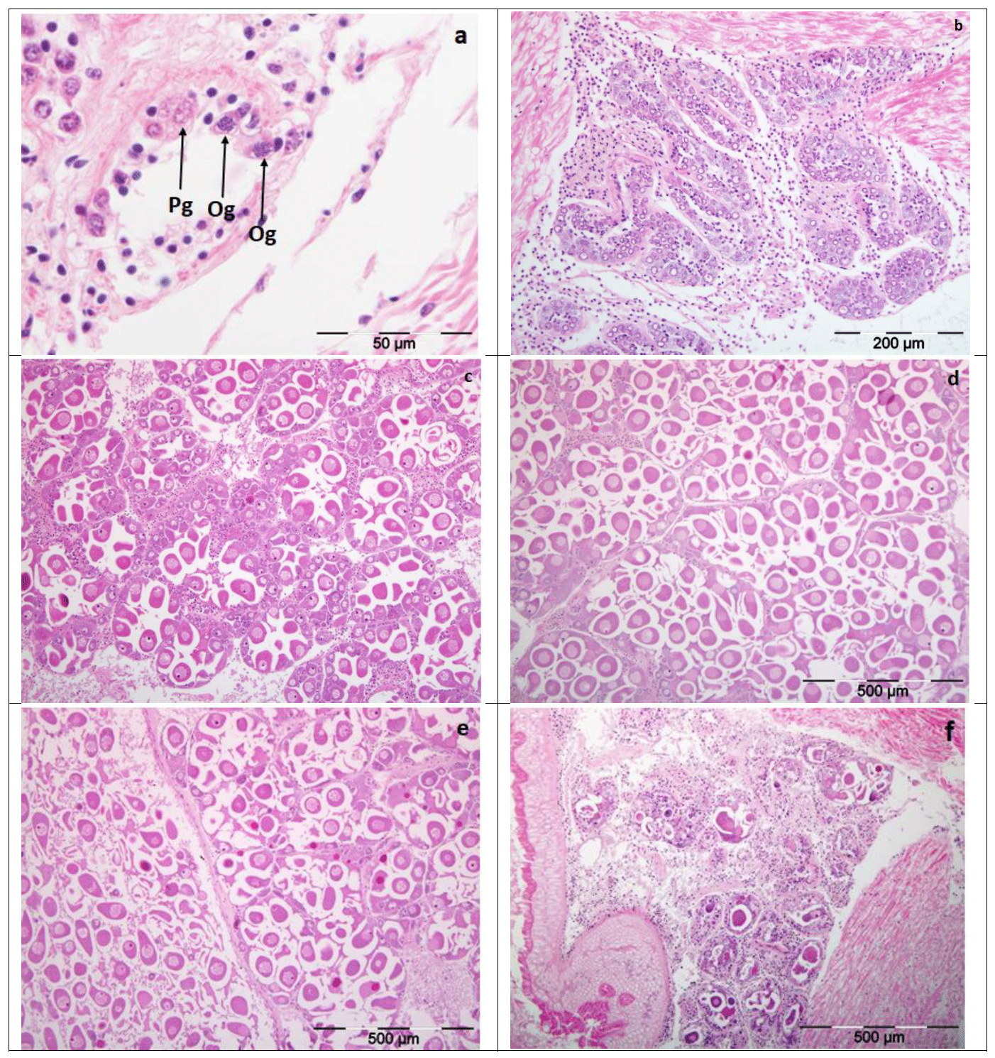
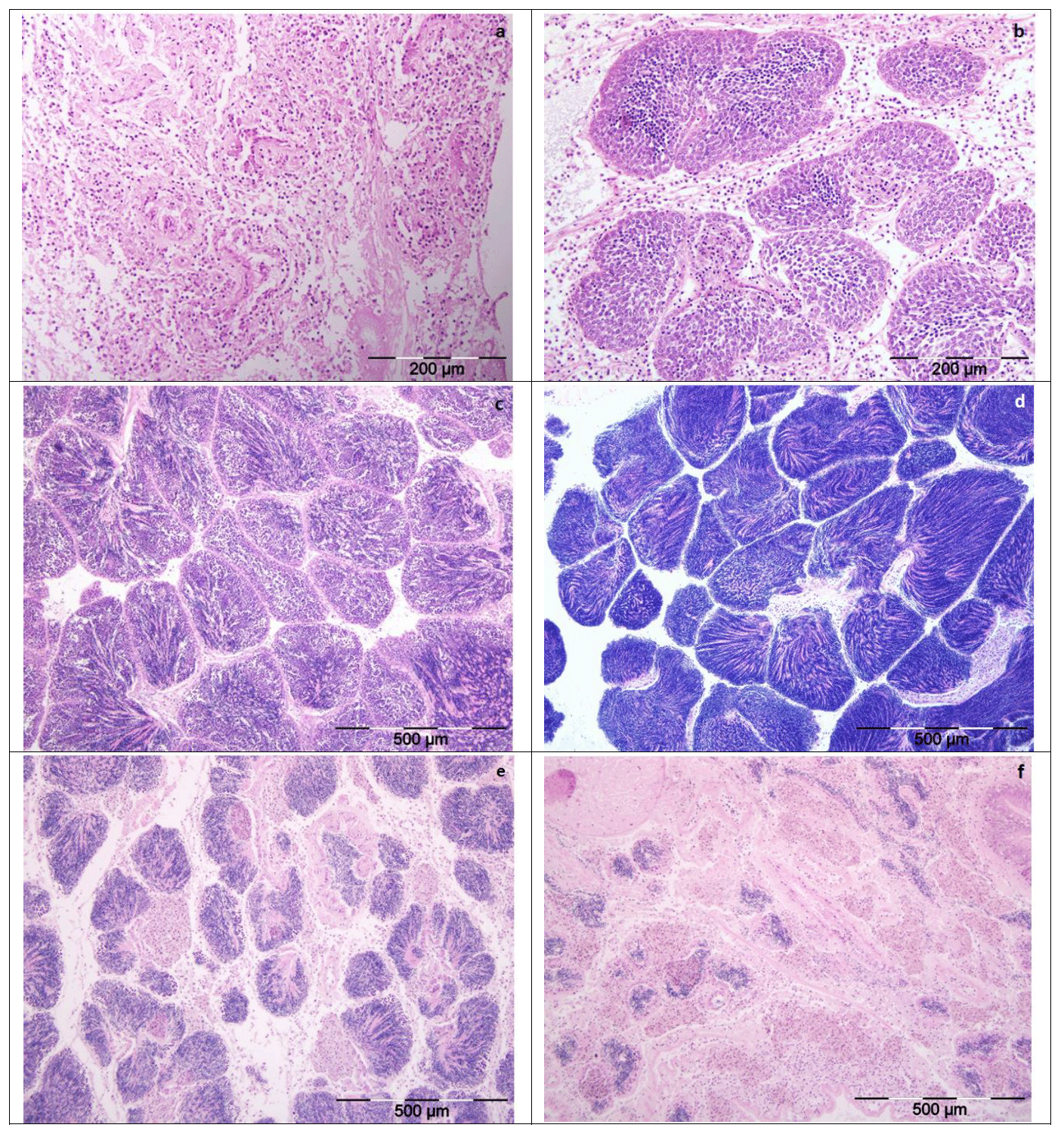
The examination of histological sections of razor clam gonad identified six different stages (0, I, II, IIIA, IIIB, IV) of gonad development (Darriba et al., 2005, Gaspar and Monteiro 1998) (Figure A6.1 and A6.2). At sexual rest (Stage 0) the gonad exhibits very few follicles, and these were small in size. Follicles also displayed protogonia and gonia in mitosis. Males exhibited marked inflammation within the follicles and the sex is clearly distinguishable. During the start of gametogenesis (Stage I), the follicle increases in size and the initial stages of gametogenesis are present. Female oocytes and previtellogenic lie at the periphery of the follicle walls (Figure A6.1b). In males, spermatogonia and spermatocytes proliferate toward the lumen (Figure A6.2b).
Advanced gametogenesis (stage II) is characterised by increased follicles which occupy the entire tissue. Germinal cells are present in all phases of gametogenesis (Figure A6.1c and A6.2c). Gonads are classified as ripe (Stage IIIA) when connective tissue has been replaced by follicles with mature gametes. The follicles exhibit a polygonal shape and are almost full of mature gametes (Figure A6.1d and A6.2d). Female oocytes display an oval or polygonal shape (Figure A6.1d) and in male the follicle lumen is full of mature spermatozoa (Figure A6.2d).
At the start of spawning (stage IIIB) the follicles start discharging. Follicle size decreases and they appear more or less empty depending on the degree of spawning (Figure A6.1e and A6.2e). Exhaustion (stage IV) is characterised by follicles small in size and almost empty. Occasional residual gametes (oocytes and spermatozoa) may be present but there is an increase in the interfollicular connective tissue (Figure A6.1f and A6.2f).
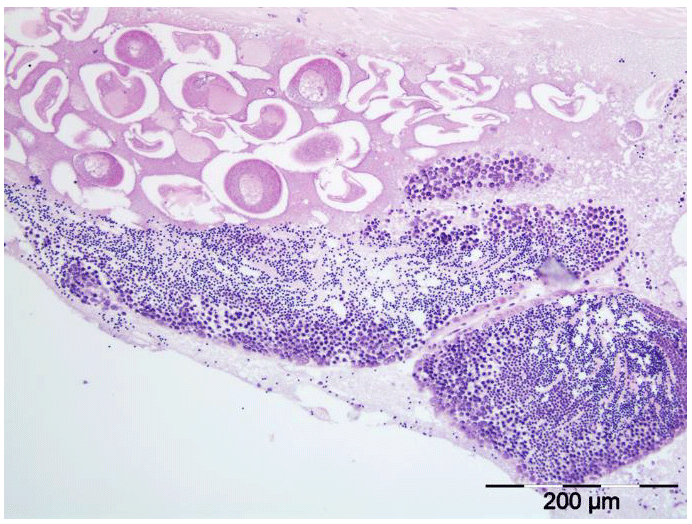
Three hermaphrodites were identified from the razor clam histology samples (one in 2020, one in 2021, and one in 2022), where both male and female gametes were observed (Figure A6.3).
Contact
There is a problem
Thanks for your feedback